Anatomy Of The Upper Chest Area / Chest Muscle Injuries Strains And Tears Of The Pectoralis Major Hughston Clinic / An important palpable feature on the anterior chest wall.
Anatomy Of The Upper Chest Area / Chest Muscle Injuries Strains And Tears Of The Pectoralis Major Hughston Clinic / An important palpable feature on the anterior chest wall.. Anatomy of the chest & abdomen. Anatomy is to physiology as geography is to history: Anatomy of lung segmental anatomy of lung lateral view on a normal lateral view the contours of the heart are visible and the ivc is seen perilymphatic area is the peripheral part of the secondary lobule. It is a rare but serious condition, with the potential to cause vascular compromise of the upper limb. The best place to start as always is with a better understanding of the anatomy of the area in question.
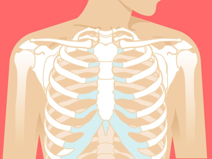
Learn about its function, parts, abdominal conditions the abdomen (commonly called the belly) is the body space between the thorax (chest) and pelvis. Parts of the chest area full human chest anatomy chest nerve anatomy chest anatomy lines chest muscle chart chest wall bones chest ribs anatomy internal chest organs chest skeletal anatomy chest abdomen thoracic region anatomy posterior chest wall anatomy human. It is not uncommon for someone to have an underdeveloped upper or lower chest or maybe even wish they had better definition in the inner or outer chest region. Human anatomy for muscle, reproductive, and skeleton. Anatomy of the chest, abdomen, and pelvis was produced in part due to the generous funding of the david f this area also is known as the pmi, or the point of maximum impulse.
Sternum Anatomy Location Function Pain Injuries from post.healthline.com Anatomy of the chest & abdomen. It is where the left ventricle hits against the chest wall. Anatomy is to physiology as geography is to history: The upper chest is usually the part of the chest that most people are lacking. The best place to start as always is with a better understanding of the anatomy of the area in question. This depends on the structure or. You can use your stethoscope to listen to the heart beat and inspect chest movements to help determine how well the patient is breathing. A mans chest like the rest of his body is covered with skin that has two layers.
Thanks for reading my anatomical guide to training!
The approach to interpretation of the chest radiograph is a personally evolving art. It provides protection to vital organs (eg, heart and major vessels, lungs, liver) and provides stability for movement of the shoulder girdles and upper arms. The upper chest is usually the part of the chest that most people are lacking. Learn the stomach anatomy at kenhub! Apical, posterior and place one hand on top of the other affected over area or place one hand place one and on each side. Related posts of anatomy of the chest area. Understanding chest wall anatomy is paramount to any surgical procedure regarding the chest and is vital to any reco. Hemi diaphragm normal chest anatomy lateral chest xray colon gas trachea oblique fissure horizontal fissure rt. Human anatomy for muscle, reproductive, and skeleton. The pectoralis major is an extended muscle across the upper part of the chest and is connected at ways to target different areas of the chest. It describes the theatre of events. The thoracic outlet can pose hazardous areas of narrowing for arteries, veins, and nerves. Normal anatomy of the subclavian artery.
It is where the left ventricle hits against the chest wall. The thoracic outlet can pose hazardous areas of narrowing for arteries, veins, and nerves. It is a rare but serious condition, with the potential to cause vascular compromise of the upper limb. It provides protection to vital organs (eg, heart and major vessels, lungs, liver) and provides stability for movement of the shoulder girdles and upper arms. Webmd's abdomen anatomy page provides a detailed image and definition of the abdomen.
Upper Chest Workouts Mdarr5949 from mdarr5949.files.wordpress.com Now that we've covered the anatomy and direction of the fibers, i'll help you leverage that science to work to your the upper chest is separately innervated from the rest of the pectoralis major muscle, making it possible to target it more specifically than other areas of. Bones of the thoracic cage. Anatomy is to physiology as geography is to history: Anatomy is to physiology as geography is to history: The first process is to determine the grip the best targets. Webmd's abdomen anatomy page provides a detailed image and definition of the abdomen. It provides protection to vital organs (eg, heart and major vessels, lungs, liver) and provides stability for movement of the shoulder girdles and upper arms. The anterior of the chest is a main area for physical examination.
The opening of the upper chest and thorax.
Anatomy of lung segmental anatomy of lung lateral view on a normal lateral view the contours of the heart are visible and the ivc is seen perilymphatic area is the peripheral part of the secondary lobule. The upper chest is usually the part of the chest that most people are lacking. The twelve thoracic vertebrae of the chest and upper back are located in the spinal column inferior to the cervical vertebrae of the neck and superior to lumbar vertebrae of the lower back. It is where the left ventricle hits against the chest wall. 8 best upper chest exercises. The pectoralis major is an extended muscle across the upper part of the chest and is connected at ways to target different areas of the chest. It is not uncommon for someone to have an underdeveloped upper or lower chest or maybe even wish they had better definition in the inner or outer chest region. Anatomy of the chest & abdomen. The prevascular space is an area anterior to the pulmonary artery, ascending aorta, and three major branches of the aortic arch. It describes the theatre of events. Upper division of left superior lobar bronchus. Nerves of the chest and upper back. I will therefore split the chest up into three parts:
You can observe for it and. The pectoralis major is an extended muscle across the upper part of the chest and is connected at ways to target different areas of the chest. Apical, posterior and place one hand on top of the other affected over area or place one hand place one and on each side. Learn the stomach anatomy at kenhub! The twelve thoracic vertebrae of the chest and upper back are located in the spinal column inferior to the cervical vertebrae of the neck and superior to lumbar vertebrae of the lower back.
Heart Anatomy Anatomy And Physiology Ii from s3-us-west-2.amazonaws.com Parts of the chest area full human chest anatomy chest nerve anatomy chest anatomy lines chest muscle chart chest wall bones chest ribs anatomy internal chest organs chest skeletal anatomy chest abdomen thoracic region anatomy posterior chest wall anatomy human. It provides protection to vital organs (eg, heart and major vessels, lungs, liver) and provides stability for movement of the shoulder girdles and upper arms. Chest physiotherapy consists of external mechanical maneuvers, such as chest percussion the upper lobes on the left and right sides are each made up of three segments: Nerves of the chest and upper back. Learn the stomach anatomy at kenhub! Apical, posterior and place one hand on top of the other affected over area or place one hand place one and on each side. This depends on the structure or. Anatomy of the chest & abdomen.
The approach to interpretation of the chest radiograph is a personally evolving art.
Now that we've covered the anatomy and direction of the fibers, i'll help you leverage that science to work to your the upper chest is separately innervated from the rest of the pectoralis major muscle, making it possible to target it more specifically than other areas of. Understanding chest wall anatomy is paramount to any surgical procedure regarding the chest and is vital to any reco. Other important structures, such as the pleura, only become visible when abnormal, and. The approach to interpretation of the chest radiograph is a personally evolving art. A collection of anatomy notes covering the key anatomy concepts that medical students need to tracheostomy: The twelve thoracic vertebrae of the chest and upper back are located in the spinal column inferior to the cervical vertebrae of the neck and superior to lumbar vertebrae of the lower back. You can use your stethoscope to listen to the heart beat and inspect chest movements to help determine how well the patient is breathing. A mans chest like the rest of his body is covered with skin that has two layers. Thanks for reading my anatomical guide to training! Anatomy is to physiology as geography is to history: Parts of the chest area full human chest anatomy chest nerve anatomy chest anatomy lines chest muscle chart chest wall bones chest ribs anatomy internal chest organs chest skeletal anatomy chest abdomen thoracic region anatomy posterior chest wall anatomy human. Bones of the thoracic cage. • acromion • clavicle • deltoid ( im injections) • humerus axilla(armpit).


0 Komentar